Introduction
Breast cancer is an ongoing concern and one of the most common forms of cancer in women. In 2018 the number was around 2,66, 120, new diagnoses in the United States alone. Invasive Ductal Carcinoma or IDC in short is one of the most common forms of breast cancer. The cancer starts in the milk duct of the breast and invades the surrounding tissue. This form of cancer makes up around 80 percent of all breast cancer diagnosis, with more than 1,80,000 women a year in the United States alone being diagnosed with IDC, according to the American Cancer Society.
The use of Artificial Intelligence can drastically reduce the need for medical staff to examine mammography slides manually, saving not only time, but money, and ultimately lives. This blog would actually showcase how the combined power of AI & IoT can accelerate the detection of IDC at a very early stage.
We all know how important the study of Histology Images can be in the analysis of microanatomy of cells, tissues and organs as seen through, with the help of an electronic microscope. This typically involves a pathological study of the tissue specimen important for biopsy, also known as a histopathology report. Moreover, it examines the correlation between structure of the cells. The contrast, colour & sharpness data of each image can be accessed by the use of USB based electronic microscope. Computer Vision or various Image Processing Techniques can very well be applied in such situations to facilitate the Diagnosis Process. This blog, Histology Data Processing & Analysis in Pathology Department, shows how existing Deep Learning Technologies can be utilized to train Artificial Intelligence (AI) to be able to detect Invasive
Ductal Carcinoma (IDC or Breast Cancer) in unlabelled Histology Images. More specifically, this blog would present the use of a Deep Learning Framework, Transfer Learning and Hyperparameter Tuning, using a Dataset of Negative and Positive Histology images using any open source or commercial cloud. In addition to presenting how artificial intelligence can be used to detect IDC, this blog would also showcase the use of Internet of Things (IoT) in conjunction with AI in order to create an automated system that can be leveraged in the medical industry as part of a future directive for accurate and expedited way of Pathological Report based Diagnostic Procedure.
Objectives & Motivation
The various Objectives & Motivations for this blog are as follows:
• Detecting Invasive Ductal Carcinoma (Breast Cancer).
• Artificial intelligence (AI) to be able to detect Invasive Ductal Carcinoma (IDC) in unlabelled Histology Images.
• Training the Dataset of Negative and Positive Histology Images using any Open Source or Commercial Cloud.
• Internet of Things (IoT) can be used as part of the Inference on Edge in conjunction with AI, to create an Automated System that can be used in the Medical Industry.
Implementation
The Cell Count via structures can be addressed by use of Morphological Operators. OpenCV supports morphological operations such as erosion, dilation, opening & closing through use of various functions. The other features like contrast and colour can be done by use of various Sharpening Filters & Colour based Processing in various planes such as L*a*b*, HSV or RGB can also be implemented to get more insight from the Histology Image. The OpenCV can also be installed in Raspberry Pi very easily. Raspberry Pi is a low power computer for Real Time Processing and well known for analysis of Images and Signals. It has an inbuilt USB interface which can be plugged in to
the Electronic Microscope via USB cable and a small API can help in pulling the Histology Image Data inside Raspberry Pi. Since the algorithms use an open-source environment for execution, Raspberry Pi board also being an open-source board provides the right platform for validating the algorithms on a real time basis. The Raspberry Pi has an HDMI port, which can be further connected to an LED
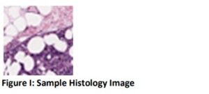
Monitor for the visualization aspect. Thus, providing an end-to-end pipeline from capturing, analysing & diagnosis. A sample Histology Image is represented in Figure I.

To introspect on the use of advanced Machine Learning Frameworks, let’s go through the following nodal points pivotal in designing of the AI Backbone for such a sensitive Bio-medical device.
a. Deep Learning Network
Convolutional neural networks are a type Deep Learning Frameworks. These types of neural nets are widely used in computer vision and have pushed the capabilities of computer vision over the last few years since their inception and have made quite progressive inroads since. It has enabled performance which would have been exceptionally difficult to be achieved by some of its previous conventional Machine
Learning peers or more traditional Neural Networks; however, studies shows that there are trade-offs related to Training times and Accuracy. In other words, it’s a non-inferior solution-based model, where you need to consider compromising on sacrificing on one
aspect i.e., Training Time or Accuracy in order to get an improvement on the other.
b. Transfer Learning
Transfer learning allows anyone to retrain the final layer of an existing model, resulting in a significant decrease in not only the Training time, but also the size of the Dataset required. One of the most famous models that can be used for transfer learning is the
Inception V3 model created by Google. This model has been trained on over a million images from 1,000 classes from the original ImageNet Dataset, which was actually trained on over 1 million Training images, the TensorFlow version has one class more viz-a-viz 1,001 classes which is due to an additional “Background’ Class not used in the original ImageNet. Being able to retrain the final layer means that you can maintain the knowledge that the model had learnt during its original training and apply it to your smaller dataset, resulting in a Highly Accurate Classification situation without the need for Extensive Training and Computational Power. In one of the previous versions, the
this model viz-a-viz an Inception V3 was retrained using transfer learning on a Raspberry Pi 3 device, so that should give you some idea of the capabilities of Transfer Learning. Some of the other important components forming a part of this ecosystem which is expected to work in conjunction with the above to achieve some of the other farreaching goals, have been listed below:
c. Cloud
Commercial Cloud or Open-source Cloud is a platform for Training Machine Learning and Deep Learning Models. Such a platform is made up of a Cluster of Servers comprising of Scalable Processors. The platform should ideally provide a number of frameworks and
tools including TensorFlow, Caffe, Keras, and Theano, as well as the Distribution for Python, required for the Deployment aspect. The Cloud is great for people getting started with learning how to Train Machine Learning and Deep Learning Models, as Graphics Processing Units (GPUs) can be quite expensive.
d. Processor for Use during Real Time Deployment
The Intel® Movidius™ Neural Compute Stick is a fairly new piece of Hardware used for enhancing the inference process of computer vision models on low-powered edge devices. The Intel Movidius product is a USB appliance that can be plugged into devices such as Raspberry Pi and basically takes the processing power off the device and onto the Intel Movidius brand chip, making the classification process a lot faster. Developers can train their models using their existing TensorFlow and Caffe scripts and, by installing the Intel Movidius Neural Compute Stick SDK on their development machine, can compile a graph that is compatible with the Intel Movidius product. A less-bulky API can
be installed on the lower-powered device allowing inference to be carried out via the Intel Movidius product. This process actually acts as an enabler to the Inference on the Edge making it useful in such sensitive Bio-medical applications
e. IoT Connectivity
The Business use case we have discussed here is an IoT enabled Alarm Device built on a Raspberry Pi device. The results that are captured from the classification of images sent to the server trigger actions on the IoT that communicate with the Raspberry Pi device.
In this case, the actions are turning on a red LED and a buzzer when Cancer is detected, and turning on a blue LED when the classification results in no cancer being detected. Obviously, this is a very simple Proof of Concept, but it shows a possibility to be
extended to more powerful applications that can save time for medical staff and, hopefully, in the right hands could help save lives through early and accurate detection.
f. Outcome: Reducing False Negatives
During testing on a larger dataset, it was noticed that misclassifications were happening resulting in False Negatives, and that in most cases the False Negatives had images in the opposite class that were very similar Prioritizing an objective depending on the application is always of paramount importance when it comes to Machine Learning. Hence, for Bio-medical applications the primary objective for any Scientist would be to Reduce the False Negatives. It’s the same for this Invasive Ductal Carcinoma Classifier as well. This stage of the project was based around tricking the classifier with very similar images from opposite classes, producing false negatives, and finding a way to be able to detect these images in advance, marking them as needing further (human) inspection. Though not a very full proof concrete approach but can be useful.
Future Directions
The use of Machine Learning / Deep Learning Techniques should not be only be limited by scope from a Computer Vision perspective. We can always open the horizon by peeping into some of the other applications where on adoption of this state-of-the-art
stuff can be a shot in the arm in the field of Medical Science going forward. For e.g.
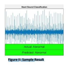
Heart Sound Classification (Figure II)
Motivation
• Heart sounds are complex signals.
• Require Medical Practitioners for Diagnosis.
Goal
• Build a Classifier and Deploy in Portable Device
Data
• Heart sound recordings (phonocardiogram).

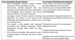
To add-on further, if we look into the spectrum of applications from two organizations
where hard-core research practices have paved way to more value-added research leading
to product development include

Name of Author: DEBAJIT SEN
E-mail id: sen.debajit@gmail.com
debajit_sen@outlook.com
Author Biography:
DEBAJIT SEN has close to 12 years’ of experience in the field of Robotic Process Automation, Artificial Intelligence & Data Science, Machine
& Deep Learning, Signal & Image Processing and Computer Vision applications. He has implemented numerous projects in the space of Manufacturing, Security & Surveillance and Biomedical & Healthcare Domain. Currently working as a Data Scientist in a Healthcare Organization. Presently, working on the development of RPA based Intelligent Content Processing (OCR & NLP based) for
Healthcare Insurance Analytics encompassing automation of Claim Forms Processing, Electronic Health Record Processing, Prior-Authorization, Dispute and Appeals & Grievances. He has worked as an Independent Researcher / Consultant collaborating for various R & D Projects with different Institutes. He has also published numerous papers in various National & International Journals,
Conferences of repute & also has an International Innovation Patent Grant from the Australian Government (IP Australia) to his name for a novel work in the domain of medical science.


